


“First-class Medical Staff & comprehensive System”
Siloam Eye Hospital will Safeguard your Health.

What is Retinal Detachment?
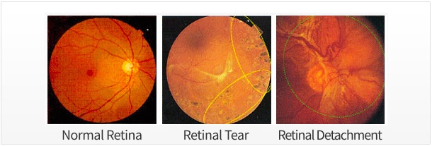
A retinal detachment is an emergency situation where a thin layer of tissue (the retina) at the back of one’s eye pulls away from its normal position. The three types of Retinal Detachment are:
Rhegmatogenous Retinal Detachment: This is the most common form of retinal detachment that occurs when there is a small tear or break in the retina. When the retina has a tear or break, the vitreous (a gel-like fluid in the center of your eye) can fill up behind the retina. The vitreous then pushes the retina away from the back of your eye, causing it to detach.
Tractional Retinal Detachment: This form of retinal detachment occurs when scar tissue on the retina pulls the retina away from the back of your eye. The most common cause of tractional retinal detachment is diabetic retinopathy, an eye condition in people with diabetes.
Exudative Retinal Detachment: This form occurs when fluid builds up behind the retina, even though there are no tears or breaks in the retina. The retina is pushed away from the back of the eye by trapped fluid, causing it to detach.
If a retinal tear is left untreated it can often progress to a stage causing retinal detachment, and advanced retinal tearing can result in the retina becoming completely separated from the optic nerve wall due to fluid filling up.
Causes of Retinal Detachment
Most cases of retinal detachment are caused by retinal tears in one or more areas.
The vitreous is a transparent, gel-like fluid that fills the center of the eyeball, occupying three-quarters of the eye with a volume of about 3.9cc. As the vitreous strongly adheres to the retina, it can pull the retina when the vitreous contracts, causing retinal tears. The contraction of the vitreous fluid is a natural occurrence with age and usually does not cause abnormalities in the retina. In most cases of retinal detachment, changes in the vitreous body is a precursor to this condition, as fluid in the vitreous flows into the lower retina when retinal tearing occurs, leading to its detachment. This detached retina fails to function properly, resulting in blurred vision and partial blindness, like a curtain suddenly dropping down.
Additionally, retinal detachment can occur as a result of severe inflammation, eye tumor complications, or diabetic retinopathy. Since this form of retinal detachment occurs without retinal tearing, it is best to treat the cause of retinal detachment and restore the retina to its normal position.
Finally, it is common for middle-aged and elderly people to experience sudden flashes (Scintillation) or see black dots floating in front of their eyes (‘Eye Floaters’)—when these symptoms occur suddenly, it is not uncommon for the contraction of vitreous to cause a tear in the retina. If one experiences these symptoms, it is vital to get a medical checkup on the condition of the vitreous and retina, because if detected early the retinal tear can be treated with Laser Photocoagulation or Cold Coagulation without the need for major surgery.
Treatment of Retinal Detachment
Special ophthalmological examinations required before hospitalization for treatment are—a Retinal Function Test, Ultrasound Examination, and Fundus Imaging.
Additionally blood tests, urine tests, chest X-rays, electrocardiograms, and lung function tests are administered to examine the patient’s general physical condition and ensure no problems may occur due to anesthesia.
In cases of Tractional Retinal Detachment caused by diabetes, a vitrectomy can remove the vitreous and scar tissue from the retina. In cases of Rhegmatogenous retinal detachment (with a hiatus hernia), surgery is required and the operation is recommended as soon as possible. In these cases it is better to stabilize the condition before the operation to prevent further retinal detachment, and any eyestrain (such as focused reading) should be avoided as much as possible.
There are additional non-invasive procedures such as Scleral Puncture and Cryotherapy that can treat the retina, as well as the invasive procedure of injecting gas or silicone oil to adhere the retina after a vitrectomy is performed to remove scar tissue. These methods may be used alone or in combination, however, injection of gas or silicon oil requires the patient to remain bedridden—laying on their stomach or side— for an extended period of a month,.